Diagnose Varicose Veins
Home / Diagnose Varicose Veins

Diagnosis
How to diagnose varicose veins?
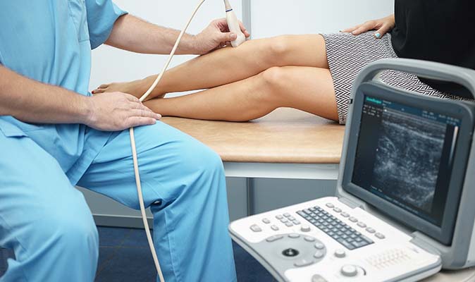
Since most veins lie deep to the skin’s surface, vein disorders are not always visible to the naked eye. This is more so in dark skinned individuals. As a result, diagnostic ultrasound (Colour Doppler) is often used to determine the cause and severity of the problem.
A MR venography may also be needed to define the veins well or look for vascular malformations. Sometimes, your surgeon may ask for an abdominal ultrasound as well, to look for any abdominal mass that may be pressing on your big veins, resulting in reflux and varicose veins. This is also useful to pick up reflux in the pelvic veins, causing pelvic congestion syndrome. Rarely, you need further tests if your surgeon feels necessary.
Classification
How to classify varicose veins?
Varicose veins are usually graded according to the CEAP classification. This included 4 parameters: Clinical, Etiology, Anatomy and Pathophysiology.
Clinical
- • C0 - No clinical signs
- • C1 - Telangiectasia(spider veins) or Reticular veins
- • C2 - Large varicose veins
- • C3 - Edema
- • C4 - Skin changes without ulceration
- • C5 - Skin changes with healed ulceration
- • C6 - Skin changes with active ulceration
Etiology
- • EC - Congenital
- • EP - Primary
- • ES - Secondary (usually our to DVT)
Anatomy
- • AS - Superficial veins
- • AD - Deep veins
- • AP - Perforating veins
Pathology
- • PR - Reflux
- • PO - Obstruction
- • PR-O - Both
When a person suffers from varicose veins he/she needs proper treatment from the experienced surgion in the field of endo venous surgery, so that they can get rid of the discomfort caused by varicose veins easily. Over the last few years, there has been a large amount of good research in the field of veins and venous diseases. Much of this has been due to the presence of sophisticated imaging technology in the form of Duplex Ultrasound, MRI and CT scan that allowed us for the first time to look inside the venous system of our body and see how they are functioning. This research was challenging and has clarified many things regarding Varicose veins disease and their treatments.